3D-Printed Facial Shields Improve Skin Cancer Treatment
Technology 'trifecta' of improved patient experience, reduced procedure time and increased accuracy
ChristianaCare’s Helen F. Graham Cancer Center & Research Institute has pioneered a new technique that significantly improves the treatment of skin cancers on the face by utilizing 3D printing technology to create custom facial shields.
This innovation, published in Practical Radiation Oncology in December 2024, has been shown to enhance patient care by increasing treatment accuracy, reducing procedure time and improving the overall patient experience.

Skin cancers, especially those on the face, can be challenging to treat due to the need for precise radiation targeting without affecting surrounding healthy tissue. Traditionally, radiation therapy for superficial skin cancers has used electron beam therapy and custom lead shields created from gypsum imprints of the patient’s face. However, this method was time-consuming and labor-intensive, often taking over 50 hours to complete.
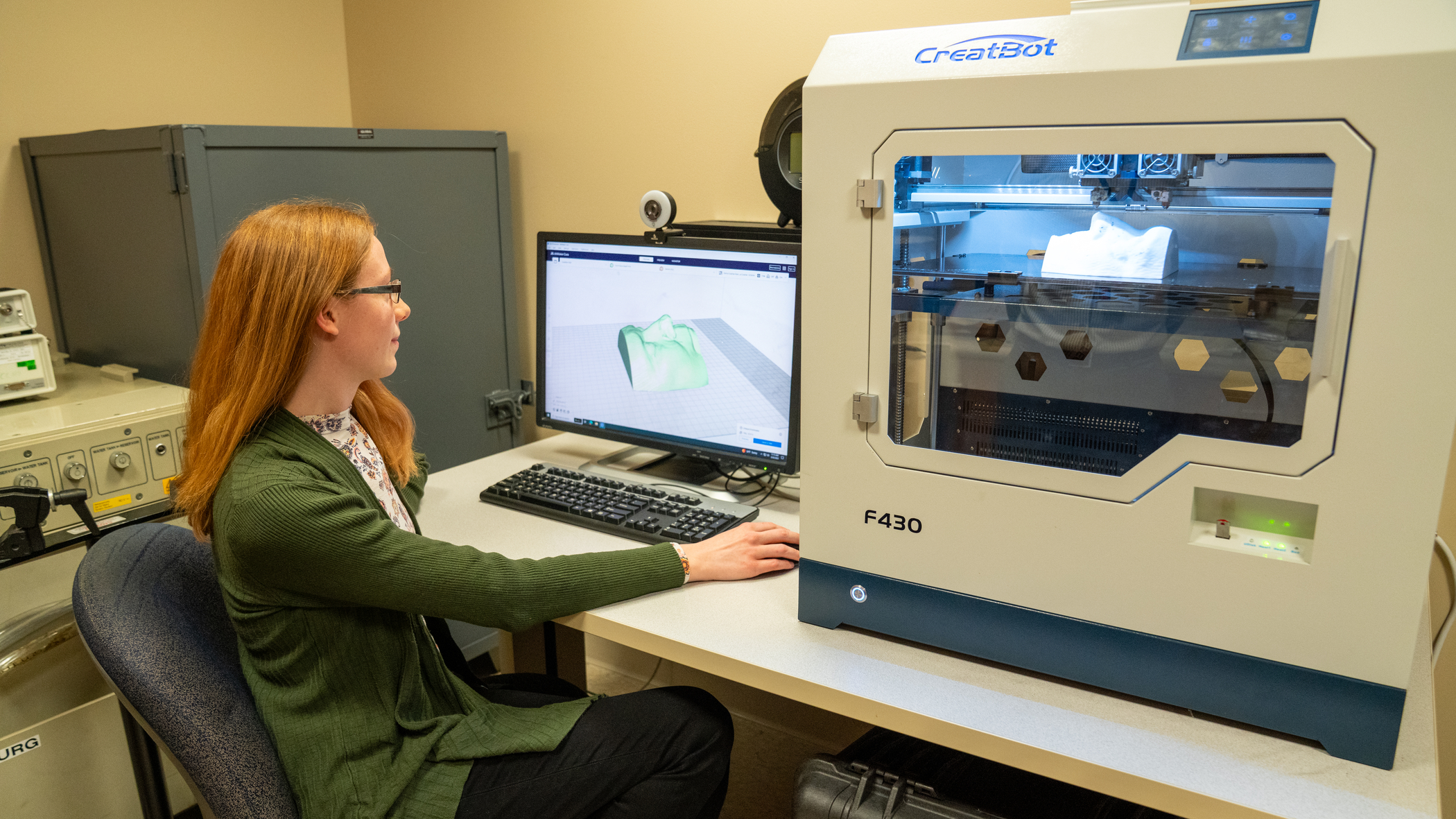
The role of 3D-printing in cancer treatment
The team at the Graham Cancer Center, including medical physicists, radiation therapists and radiation oncologists, introduced a cutting-edge alternative: 3D-printed custom facial shields. By using 3D printing technology, the team can now create highly accurate, patient-specific facial models that precisely match each patient’s anatomy, improving both the efficiency and precision of treatment.
Click here to make an appointment at the Graham Cancer Center.
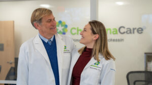
“We hit the trifecta with this new method – improving the patient experience, reducing procedure and labor time and increasing accuracy,” said Hank Chen, MS, senior medical physicist at the Graham Cancer Center.
“This approach allows us to deliver radiation to a very specific area of the face while minimizing exposure to surrounding tissues, which is critical in such sensitive areas. It’s faster, more accurate, and provides a more comfortable experience for our patients.”

The study compared the traditional gypsum-imprinted model (GIM) with the new 3D-printed custom model (3D-PCM) for patients requiring electron therapy aimed directly to the nose.
The results showed that the 3D-PCM not only provided a higher degree of anatomical accuracy but also required significantly less time to create — just 6.5 hours, compared to 54.5 hours for the GIM.
Additionally, the 3D-PCM process is less labor-intensive and improves patient comfort by offering a more effective fit.
Setting a new standard in skin cancer care

While the 3D-PCM technique does require CT scanning to capture patient anatomy, the team noted that the radiation exposure from the CT scan is minimal, and future developments, such as 3D-camera scanning, may completely eliminate the need for CT imaging.
This advancement has become standard practice for the treatment of skin cancers at the Graham Cancer Center, and the innovation is expected to set a new benchmark in skin cancer care, making it more personalized and accessible to patients.
“By incorporating 3D printing into our treatment process, we’re able to create a more efficient workflow and provide a higher level of care for our patients,” said Laura Doyle, Ph.D., DABR, chief clinical physicist at the Graham Cancer Center.
“This technology represents a significant leap forward in the field of radiation oncology, and we’re proud to be at the forefront of its application.”