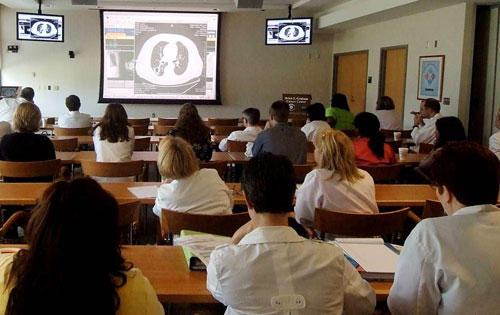
Treating lung cancer: A multidisciplinary team approach


Lung cancer is the third most commonly diagnosed cancer in Delaware.
Patients who choose an experienced cancer center with a qualified team of specialists broaden treatment options and potentially improve outcomes. In 2009, the Delaware Cancer Registry recorded 779 newly diagnosed/treated cases of lung cancer. More than half (413), from Delaware and out of state, were diagnosed or received initial treatment at the Helen F. Graham Cancer Center at Christiana Care.
Prevalence of lung cancer
According to the latest figures from the Delaware Division of Public Health, lung cancer accounts for 16 percent of all new cancer cases. Incidence increases with age, peaking at ages 75-84.
Although more men than women get lung cancer, the incidence for Caucasian women is rising. Lung cancer is the leading cause of death among Delaware men and women, accounting for 31.1 percent of all cancer deaths.
Historically, the rate of lung-cancer deaths among Delawareans was much higher than the national average, but that gap is closing. The rate of decline, however, is much faster for men than for women.
Preventing lung cancer
Smoking is the biggest risk factor for lung cancer. Other risks include exposure to environmental or workplace contaminants such as asbestos or radon.
Tobacco use causes an estimated 87 percent of all lung cancer cases. Nonsmokers who breathe the smoke of others also risk developing lung cancer. The best way to prevent lung cancer is to avoid tobacco smoke. The Helen F. Graham Cancer Center offers patients, family members and other caregivers help to quit smoking. (Two-year funding comes from the American Recovery and Investment Act through the NCI Community Cancer Centers Program.) Face-to-face and telephone sessions with a specially trained smoking-cessation counselor can identify individual smoking triggers and develop strategies to kick the habit. The counselor also evaluates health problems related to ending nicotine addiction and the effectiveness of quitting with nicotine-replacement products.
The Smoking Cessation line offers more information at 302-623-4443.
Signs and symptoms
People with lung cancer can experience fatigue, shortness of breath, chest pain, loss of appetite or persistent coughing, and bringing up mucus or blood. In some cases, a chest X-ray or CT scan performed for some other reason reveals lung cancer.
Many people do not experience symptoms until the tumor starts to grow or the cancer begins to spread to other parts of the body. This can cause worsening breathing difficulties, bone pain, stomach or back pain, headache, weakness, seizures and speech problems.
Some lung cancers can cause a group of symptoms described as syndromes. Rarely, a lung tumor can cause chemical imbalances detected in the blood.
Types of lung cancer
Lung cancer most often starts in the cells that line the airways and produce mucus. As the cancer grows, the cells can break away and spread to the lymph nodes and other parts of the body. Lung cancer is life-threatening because it often begins to spread before it is found. Non-small-cell lung cancer is the most common type. There are three main sub-types in this group:
- Squamous cell carcinoma accounts for about 25 to 30 percent of lung cancers. They occur usually in the middle of the lung and are linked to smoking.
- Adenocarcinoma accounts for about 40 percent of lung cancers, found in the outer part of the lung. This type of cancer occurs more often in young people, mainly those who smoke, but it is also the most common type of lung cancer for nonsmokers.
- Large-cell (undifferentiated) carcinoma accounts for about 10 to 15 percent of lung cancers, can start in any part of the lung and can spread quickly, making it harder to treat.
Only about 10 to 15 percent of all lung-cancer cases are small-cell lung cancer, which usually starts in the breathing tubes (bronchi) in the center of the chest. This type of lung cancer can grow quickly into large tumors and spread rapidly to other parts of the body.
Diagnosing lung cancer
The work-up for diagnosing lung cancer starts with a detailed medical history and physical exam. The doctor orders imaging tests, including chest x-ray, CT scan, MRI, PET scan or bone scan. If any of these tests suggests the possibility of lung cancer, the pathologist diagnoses lung cancer by examining a biopsied tissue sample under the microscope.
Staging
A process called staging determines the extent and size of the lung cancer and how widely it has spread. Staging uses a system developed by the American Joint Committee on Cancer (AJCC) and provides information for determining treatment and predicting survival.
| Stages of non-small-cell lung cancer | |
|---|---|
| Occult stage | Cancer cells are present in the sputum or other lung fluids, but no detectable lung tumors. |
| Stage 0 | Cancer is localized and has not spread beyond the inner lining of the lung. |
| Stage I | Cancer is small and has not spread to the lymph nodes. |
| Stage II | Cancer has grown larger, possibly into nearby structures and may have spread to nearby lymph nodes. |
| Stage III | Cancer has spread to the chest wall or diaphragm; or to lymph nodes between the lungs; or far away lymph nodes in the chest or neck. |
| Stage IV | Cancer has spread to other parts of the body, such as the other lung, brain, bones or liver. Small-cell lung cancer is classified as either limited (cancer is only in the chest) or extensive (cancer has spread outside the chest). |
Advanced, minimally invasive techniques
The Helen F. Graham Cancer Center multidisciplinary lung cancer treatment team specializes in the most advanced, minimally invasive ways to detect lung cancer early, reducing the need for higher-risk procedures and increasing treatment options. Interventional pulmonologists Gerald O’Brien, M.D., and Tuhina Raman, M.D., specialize in the latest bronchoscopy techniques, offered at few centers in the region, to diagnose and stage lung cancer, facilitate treatment and manage symptoms that may restrict breathing or cause pain.
Electromagnetic navigation bronchoscopy (ENB) works much like an automobile’s GPS system to guide placement of a flexible bronchoscope catheter deep within the lungs, following a map generated from the patient’s CT scan. Once the catheter is removed, a sheath that extends beyond the reach of the bronchoscope provides a channel to obtain tissue and fluid samples for diagnosis and staging, to facilitate pre-operative planning and minimally invasive surgical procedures or to insert markers for radiotherapy.
Surgery
Conventional surgery to remove the cancer and possibly the lung or portions of the lung may be an option for early non-small-cell lung cancer. Surgery can be combined with other treatments. For small-cell lung cancer, chemotherapy is usually the main treatment, alone or with radiation. Very rarely, surgery may be an option for very early stage small-cell lung cancer.
Video-assisted thoracic surgery (VATS) is less invasive than conventional surgery but requires a great deal of skill and experience. At the Helen F. Graham Cancer Center, thoracic surgeons Tom Bauer, M.D., and Charles Mulligan, M.D., specialize in VATS to treat early-stage lung cancer. With VATS, tiny cameras allow surgeons to view and remove tumors inside the lungs by making only a few small incisions. Surgeons use VATS most often for smaller tumors near the outside of the lung. Patients experience less pain, shorter hospital stays and quicker recovery than with conventional surgery.
Radiation and chemotherapy
Most people with lung cancer undergo more than one type of treatment. Depending on the size and stage of the cancer, doctors use chemotherapy and radiation alone or in combination as a primary treatment. These treatments can shrink a tumor before surgery or destroy remaining cancer cells after surgery.
When surgery is not an option, radiation and chemotherapy can treat invasive cancers or relieve symptoms. Advances in “personalized” therapy allow doctors to target a cancer’s specific genes, proteins or tissue environment to block growth and spread of the cancer.
At the Helen F. Graham Cancer Center, Radiation Oncology, chaired by Christopher Koprowski, M.D., employs the latest technologies to safely deliver powerful external-beam radiation to lung tumors. Precise treatment planning afforded by four-dimensional, conformal radiation, intensity-modulated radiation therapy (IMRT) and image-guided radiotherapy (IGRT) enables radiation oncologists to size and shape the radiation beam to the tumor and destroy cancer cells more effectively with low risk of treatment side effects or damage to nearby healthy tissues and organs.
CyberKnife treatment is a new, less-invasive form of radiation therapy that offers new lung cancer treatment options to patients who cannot tolerate surgery or whose tumors are inoperable. Christiana Care offered the first and only CyberKnife treatment in Delaware and one of only a few in the Mid-Atlantic region. CyberKnife uses a combination of computers, image-guided cameras and robotic technology to deliver high doses of radiation to tumors with extreme accuracy, even in difficult-to-reach areas. There is no anesthesia, no pain and minimal recovery time.
Researching new ways to fight cancer
The Helen F. Graham Cancer Center is among an elite group of research centers working with pharmaceutical companies, other research sites and the National Cancer Institute Community Clinical Oncology Program (CCOP) to streamline clinical pathways to the most promising new anti-cancer therapies.
Surviving lung cancer
Unfortunately, the ability to find lung cancer early and treat it effectively is more challenging than for some other cancers. More than 70 percent of lung cancers are diagnosed in the late stages, when treatment is much more difficult. However, according to the latest available figures (2009), patients with early-stage non-small-cell lung cancer diagnosed or treated at Christiana Care have a five-year survival rate of at least 49 percent, well above Delaware averages for the same period and the latest available (2002) national averages recorded in the National Cancer Data Base.
Numbers such as these do not always tell the whole story, because they include patients who have elected to have no treatment at all. Five-year survival rates are lower for patients with more deeply invasive lung cancer, underscoring the need for earlier screening capabilities and better treatment through clinical trials.
Multidisciplinary cancer care
At the Helen F. Graham Cancer Center, patients who have lung cancer and their loved ones meet with a team of cancer specialists, including a medical oncologist, an interventional pulmonologist, a surgeon, a radiation oncologist and a cancer nurse navigator at the Multidisciplinary Lung Cancer Center. Together they discuss the latest thinking and most promising treatments.
Recent evidence suggests that lung-cancer patients who receive palliative care early in their treatment plans increase long-term survival and experience an improved quality of life. With help from a nurse navigator, patients easily access the many palliative and support services available, including nutrition counseling, social work, wellness coaching, psychological counseling, pain and symptom management, oncology rehabilitation, genetic counseling and support groups, including cancer companion and survivorship programs.
As one of the first selected National Cancer Institute Community Cancer Centers, the Christiana Care Helen F. Graham Cancer Center is a national model for the diagnosis, treatment and prevention of cancer. For this article, six cancer specialists shared their insights into a multidisciplinary approach to diagnosing and treating lung cancer. They are Thomas L. Bauer, II, M.D., chief of Thoracic Surgery, Michael J. Guarino, M.D., medical oncologist and director of the Pharmaceutical Clinical Trials Program, Christopher Koprowski, M.D., MBA, chief of Radiation Oncology, Gregory A. Masters, M.D., medical oncologist and director of the Medical Oncology/Hematology Fellowship Training Program, Gerald M. O’Brien, M.D., director of Bronchoscopy and Interventional Pulmonology, and Robert McBride, director of the Tumor Registry.