New cardiac PET scans offer multiple advantages for coronary artery disease diagnosis


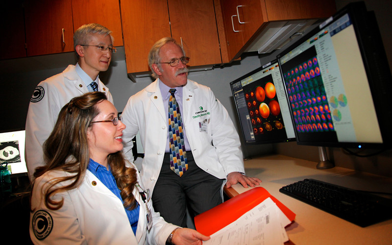
Christiana Care’s Section of Nuclear Medicine and Cardiovascular Laboratory are introducing stress myocardial perfusion Positron Emission Tomography (PET) scans, a technology that produces high-quality images that will make it easier to diagnose coronary artery disease.
The program is initially focusing on patients who are obese or women who have large breasts, conditions that make it difficult to achieve accurate results with traditional stress tests, said Anthony Gialloreto, MSHCA, CNMT, RT-N, director, Non-Invasive Services.
“We expect this technology will be a great help with these more difficult diagnoses because the resolution of the images is so much clearer,” he said.
Cardiac PET scans offer significant advantages over a traditional stress myocardial perfusion scan using Single Photon Emission Computed Tomography (SPECT). “They are more time efficient and can be completed in about an hour, compared to approximately three to four hours for SPECT studies,” said Hung Dam, M.D., associate medical director of Nuclear Medicine. “In addition, patients and the monitoring medical staff are exposed to less radiation from PET scans compared to SPECT tests.”
Clinical studies have shown that cardiac PET scans are more accurate than SPECT tests. Patients receive 82Rubidium chloride, a radioactive tracer that is injected into the bloodstream. A PET camera detects the tracer as it flows through cardiac vasculature to create high-quality pictures of the heart.
“Patients also benefit because the technology greatly reduces false positive results,” said Erik Marshall, M.D., medical director of Christiana Care’s Non-invasive Cardiology Laboratory. That is of particular concern for patients whose abundant soft tissue can make it more difficult to ensure accurate results.