
Page 15 - Demo
P. 15
| Cancer Care
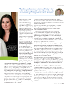
“Together, we have succeeded in achieving better
value and an improved treatment experience for
women with gynecological cancers in Delaware.”
Kai Yang, MS, Medical
Physicist II, Radiation Oncology, Lana De Souza Lawrence, M.D., Radiation Oncology, Elias Chua, M.D., Anesthesia Service, Lauren Davidson BS, RT (R) (MR), MRI section supervisor, Radiology.
Firas Mourtada, MSE, Ph.D., D.ABR
women with gynecological cancers in Delaware.”
Compared with traditional computer tomography (CT) scans, MRI offers a better view of the tumor and other critical structures to more precisely target the cancer while sparing surrounding normal tissues and organs. The Graham Cancer Center is one of only a handful of U.S. community cancer centers to incorporate MRI into its radiation oncology imaging toolbox.
With HDR brachytherapy, the radiation oncology team uses specially designed applicators placed in and adjacent to the cervix to deliver precisely
the prescribed high doses of radiation directly to the cancer cells. Before the new protocol was put in place, diagnostic MR images taken some weeks prior to treatment were available
to the team, but for treatment planning on the day of the pro- cedure, they relied on CT scan images of the tumor anatomy.
“Because we worked in parallel rather than serially,” said Dr. Mourtada, “we actually completed treatment on the first patient in less time than we would have taken with a CT-based plan, and with less stress on the patient.”
The team even worked with a local manufacturer to design an MRI-compatible transport table or “hover board” to safely assist with the transfer of the patient between procedures without compromising image quality.
“We had to work creatively and compatibly to ensure that
all devices and transfer equipment complied with the safety requirements of the unique MRI environment,” said MRI Section Supervisor Lauren Davidson, BS, RT (R) (MR), team leader for Radiology. “We were able to simulate the process step-by-step
to ensure a smooth workflow on the actual day of imaging and consequently reduce the patient’s time in the scanner down to six minutes from our planning time of 20 minutes.”
“As a hybrid academic community cancer center, we must think creatively to find practical solutions to cost and distance so that advancing technologies like MRI integrated treatment planning become accessible to our patients in their own communities,” said Nicholas Petrelli, M.D., Bank of America endowed medical director of the Helen F. Graham Cancer Center & Research Institute. “Dr. Mourtada and Dr. DeSouza Lawrence along with the rest of the team have shown that the answer is not always about spending the most dollars or buying the biggest technology, but that innovation and value can effectively drive success.”
In recent years, there has been growing interest in using MRI either alone or in conjunction with computed tomography (CT) for guided imaging. Unlike X-rays or CT scans, MRI uses no radiation. Also, MRI scans can be programmed for different (multi) physical parameters to highlight specific differences between cancer and normal tissues.
Dr. Mourtada chairs a task force for the American Association
of Physicists in Medicine, charged over the next two years with writing national standards for MRI with brachytherapy for prostate and gynecological cancers.
“There is no question that this modality is becoming the standard,” he said. “However, unlike in Europe, we in the U.S. continue to look for ways to adapt within our constraints to provide our patients the best image modality to manage their disease effectively and safely. What we are learning from our experiences here at the Graham Cancer Center, we are able to share with other centers around the country.”
FOCUS • MAY 2018 13
“The advantage of having the MR image with the applicator in place just prior to treatment
is that the cervix can be in a very different position from when a diagnostic MRI was done without the device in place,” said Lana DeSouza Lawrence, M.D., radiation oncologist at the Helen F. Graham Cancer Center & Research Institute. Her patient, a 58-year- old woman with stage IV cervical cancer, became the first patient in Delaware to benefit from the new MRI-based brachytherapy.
“With MRI we can be more precise about delineating tumor boundaries in relation to surrounding tissues and organs that we do not want to treat with radiation,” Dr. DeSouza Lawrence said.
Anesthesiologists Pam Moore, M.D., team leader for Radiation Oncology, and Elias Chua, M.D., consulted on workflow design. “The driver for us in developing this new process was patient safety and patient satisfaction,” Dr. Moore said.
